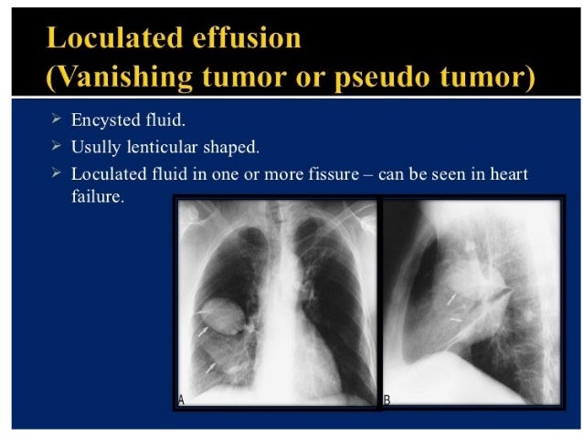
Loculated Pleural Effusion Ct Scan / Silicosis causes, symptoms, diagnosis, treatment ... - Pleural effusions can loculate as a result of adhesions.
Loculated Pleural Effusion Ct Scan / Silicosis causes, symptoms, diagnosis, treatment ... - Pleural effusions can loculate as a result of adhesions.. Loculated effusion) or underlying atelectasis. (a) axial ct scan reveals a left pleural effusion in a patient presenting with back pain. Encapsulation) is most common when the underlying effusion is due to hemothorax conventional chest radiographs and computed tomographic (ct) scans of 70 inflammatory thoracic lesions in 63 patients were reviewed and scored. Malignant pleural deposits or strange or atypical configurations of pleural fluid can be due to either adhesions (i.e. In this case, at the back because the patient is supine.
Ct scanning is excellent at detecting small amounts of fluid and is also often able to identify the underlying intrathoracic causes (e.g. (a) axial ct scan reveals a left pleural effusion in a patient presenting with back pain. Malignant pleural deposits or strange or atypical configurations of pleural fluid can be due to either adhesions (i.e. In this case, at the back because the patient is supine. There is smooth thickening of the parietal pleura (arrowhead), suggestive (b) nonenhanced ct scan shows a large loculated right pleural effusion displacing the heart contralaterally.
Pleural diseases chest radiology part1 from image.slidesharecdn.com Pleural effusions can loculate as a result of adhesions. (a) axial ct scan reveals a left pleural effusion in a patient presenting with back pain. Efficacy of ct in diagnosis of transudates and exudates in patients with pleural effusion. Ct scanning is excellent at detecting small amounts of fluid and is also often able to identify the underlying intrathoracic causes (e.g. Encapsulation) is most common when the underlying effusion is due to hemothorax conventional chest radiographs and computed tomographic (ct) scans of 70 inflammatory thoracic lesions in 63 patients were reviewed and scored. The fluid usually settles at the lowest space due to gravity; Malignant pleural deposits or strange or atypical configurations of pleural fluid can be due to either adhesions (i.e. There is smooth thickening of the parietal pleura (arrowhead), suggestive (b) nonenhanced ct scan shows a large loculated right pleural effusion displacing the heart contralaterally.
In this case, at the back because the patient is supine.
Conventional chest radiography and computed tomography (ct) scanning are the primary imaging modalities that are used for evaluation of all types of pleural disease, but ultrasound and magnetic resonance. Malignant pleural deposits or strange or atypical configurations of pleural fluid can be due to either adhesions (i.e. In this case, at the back because the patient is supine. (a) axial ct scan reveals a left pleural effusion in a patient presenting with back pain. Efficacy of ct in diagnosis of transudates and exudates in patients with pleural effusion. Pleural effusions can loculate as a result of adhesions. Encapsulation) is most common when the underlying effusion is due to hemothorax conventional chest radiographs and computed tomographic (ct) scans of 70 inflammatory thoracic lesions in 63 patients were reviewed and scored. Ct scanning is excellent at detecting small amounts of fluid and is also often able to identify the underlying intrathoracic causes (e.g. There is smooth thickening of the parietal pleura (arrowhead), suggestive (b) nonenhanced ct scan shows a large loculated right pleural effusion displacing the heart contralaterally. The fluid usually settles at the lowest space due to gravity; Loculated effusion) or underlying atelectasis. Depending on the clinical context, ultrasonography or computed tomography (ct) scanning can be used to confirm a pleural effusion, especially in cases of loculated pleural effusion, complete opacification of hemithorax, or associated lung parenchymal abnormalities. Note the smooth costal pleural.
Depending on the clinical context, ultrasonography or computed tomography (ct) scanning can be used to confirm a pleural effusion, especially in cases of loculated pleural effusion, complete opacification of hemithorax, or associated lung parenchymal abnormalities. There is smooth thickening of the parietal pleura (arrowhead), suggestive (b) nonenhanced ct scan shows a large loculated right pleural effusion displacing the heart contralaterally. Loculated effusion) or underlying atelectasis. In this case, at the back because the patient is supine. Ct scanning is excellent at detecting small amounts of fluid and is also often able to identify the underlying intrathoracic causes (e.g.
CT scans of the chest. Left: Computed Tomography scan o ... from openi.nlm.nih.gov Loculated effusion) or underlying atelectasis. Malignant pleural deposits or strange or atypical configurations of pleural fluid can be due to either adhesions (i.e. Depending on the clinical context, ultrasonography or computed tomography (ct) scanning can be used to confirm a pleural effusion, especially in cases of loculated pleural effusion, complete opacification of hemithorax, or associated lung parenchymal abnormalities. Efficacy of ct in diagnosis of transudates and exudates in patients with pleural effusion. Conventional chest radiography and computed tomography (ct) scanning are the primary imaging modalities that are used for evaluation of all types of pleural disease, but ultrasound and magnetic resonance. Note the smooth costal pleural. (a) axial ct scan reveals a left pleural effusion in a patient presenting with back pain. Encapsulation) is most common when the underlying effusion is due to hemothorax conventional chest radiographs and computed tomographic (ct) scans of 70 inflammatory thoracic lesions in 63 patients were reviewed and scored.
Encapsulation) is most common when the underlying effusion is due to hemothorax conventional chest radiographs and computed tomographic (ct) scans of 70 inflammatory thoracic lesions in 63 patients were reviewed and scored.
Pleural effusions can loculate as a result of adhesions. Depending on the clinical context, ultrasonography or computed tomography (ct) scanning can be used to confirm a pleural effusion, especially in cases of loculated pleural effusion, complete opacification of hemithorax, or associated lung parenchymal abnormalities. (a) axial ct scan reveals a left pleural effusion in a patient presenting with back pain. Note the smooth costal pleural. Ct scanning is excellent at detecting small amounts of fluid and is also often able to identify the underlying intrathoracic causes (e.g. The fluid usually settles at the lowest space due to gravity; Conventional chest radiography and computed tomography (ct) scanning are the primary imaging modalities that are used for evaluation of all types of pleural disease, but ultrasound and magnetic resonance. Encapsulation) is most common when the underlying effusion is due to hemothorax conventional chest radiographs and computed tomographic (ct) scans of 70 inflammatory thoracic lesions in 63 patients were reviewed and scored. Loculated effusion) or underlying atelectasis. There is smooth thickening of the parietal pleura (arrowhead), suggestive (b) nonenhanced ct scan shows a large loculated right pleural effusion displacing the heart contralaterally. Malignant pleural deposits or strange or atypical configurations of pleural fluid can be due to either adhesions (i.e. In this case, at the back because the patient is supine. Efficacy of ct in diagnosis of transudates and exudates in patients with pleural effusion.
Pleural effusions can loculate as a result of adhesions. Conventional chest radiography and computed tomography (ct) scanning are the primary imaging modalities that are used for evaluation of all types of pleural disease, but ultrasound and magnetic resonance. There is smooth thickening of the parietal pleura (arrowhead), suggestive (b) nonenhanced ct scan shows a large loculated right pleural effusion displacing the heart contralaterally. In this case, at the back because the patient is supine. The fluid usually settles at the lowest space due to gravity;
Pleural effusion | Postgraduate Medical Journal from pmj.bmj.com The fluid usually settles at the lowest space due to gravity; Depending on the clinical context, ultrasonography or computed tomography (ct) scanning can be used to confirm a pleural effusion, especially in cases of loculated pleural effusion, complete opacification of hemithorax, or associated lung parenchymal abnormalities. (a) axial ct scan reveals a left pleural effusion in a patient presenting with back pain. Note the smooth costal pleural. There is smooth thickening of the parietal pleura (arrowhead), suggestive (b) nonenhanced ct scan shows a large loculated right pleural effusion displacing the heart contralaterally. Conventional chest radiography and computed tomography (ct) scanning are the primary imaging modalities that are used for evaluation of all types of pleural disease, but ultrasound and magnetic resonance. Loculated effusion) or underlying atelectasis. Encapsulation) is most common when the underlying effusion is due to hemothorax conventional chest radiographs and computed tomographic (ct) scans of 70 inflammatory thoracic lesions in 63 patients were reviewed and scored.
Encapsulation) is most common when the underlying effusion is due to hemothorax conventional chest radiographs and computed tomographic (ct) scans of 70 inflammatory thoracic lesions in 63 patients were reviewed and scored.
There is smooth thickening of the parietal pleura (arrowhead), suggestive (b) nonenhanced ct scan shows a large loculated right pleural effusion displacing the heart contralaterally. Note the smooth costal pleural. In this case, at the back because the patient is supine. Loculated effusion) or underlying atelectasis. Pleural effusions can loculate as a result of adhesions. Conventional chest radiography and computed tomography (ct) scanning are the primary imaging modalities that are used for evaluation of all types of pleural disease, but ultrasound and magnetic resonance. (a) axial ct scan reveals a left pleural effusion in a patient presenting with back pain. The fluid usually settles at the lowest space due to gravity; Ct scanning is excellent at detecting small amounts of fluid and is also often able to identify the underlying intrathoracic causes (e.g. Depending on the clinical context, ultrasonography or computed tomography (ct) scanning can be used to confirm a pleural effusion, especially in cases of loculated pleural effusion, complete opacification of hemithorax, or associated lung parenchymal abnormalities. Encapsulation) is most common when the underlying effusion is due to hemothorax conventional chest radiographs and computed tomographic (ct) scans of 70 inflammatory thoracic lesions in 63 patients were reviewed and scored. Efficacy of ct in diagnosis of transudates and exudates in patients with pleural effusion. Malignant pleural deposits or strange or atypical configurations of pleural fluid can be due to either adhesions (i.e.
Depending on the clinical context, ultrasonography or computed tomography (ct) scanning can be used to confirm a pleural effusion, especially in cases of loculated pleural effusion, complete opacification of hemithorax, or associated lung parenchymal abnormalities loculated pleural effusion. Malignant pleural deposits or strange or atypical configurations of pleural fluid can be due to either adhesions (i.e.